This patient came to our attention 4 years ago for Vitelliform Macular Dystrophy or Best Disease. (See below for a description of this condition).
Initially, a layered yellow spot is visible in the right eye on retinography, and on OCT, the spot appears hyperdense in the lower part and hypodense in the upper part.
On fluorescein angiography, the yellowish part appears hypofluorescent because it masks the underlying retina, while the upper part is hyperfluorescent due to an accumulation of the dye.
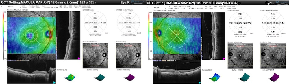
In the right eye, the yellowish material is then reabsorbed, and in its place, the retina atrophies, as is clearly evident on OCT.
In the left eye, which initially appears almost normal with little accumulation of subretinal material, there is a progressive accumulation of material over the years.

Best Vitelliform Macular Dystrophy: A Challenge to Central Vision
Best Vitelliform Macular Dystrophy (BVMD), also known as Best Disease, represents one of the most fascinating and complex challenges in contemporary ophthalmology. This genetic condition, which affects central vision, brings with it a series of symptoms and complications that require constant attention and targeted management.
A Rare Disease with a Profound Impact
BVMD, although rare, is a disease that can significantly influence patients’ quality of life. Its prevalence is estimated between 1/5,000 and 1/67,000, with a slight predisposition towards males. Symptoms typically begin during childhood or adolescence, with normal vision at birth that progressively deteriorates over time.
A Complicated Progression
BVMD goes through several stages, each bringing its own symptoms and characteristics. From the initial asymptomatic phase, it progresses to the formation of the characteristic “egg yolk” lesion in the macula, followed by the appearance of vitelliform material that can lead to scar formation and consequent deterioration of central visual acuity. The disease can be complicated by the development of a choroidal neovascular membrane, which can further worsen the visual prognosis.
Genetic Origin
BVMD is characterized by mutations in the BEST1 gene, located on chromosome 11q12, leading to the abnormal production of a protein called bestrophin-1. This anomaly results in an accumulation of lipofuscin, a substance that damages the retinal pigment epithelium and contributes to central vision dysfunction.
Comprehensive and Accurate Diagnosis is Crucial
Diagnosing BVMD requires a thorough clinical evaluation, which may include family history, fundus examination, and the use of advanced diagnostic techniques such as fluorescein angiography and OCT. The diagnosis is confirmed through genetic screening.
Treatment
The treatment of BVMD primarily focuses on symptom management and complication prevention. Therapeutic interventions may include the use of low vision aids, avoiding smoking to reduce the risk of vascular complications, and using therapies such as photodynamic therapy or anti-VEGF agents to treat the choroidal neovascular membrane.
Prognosis is Variable
The prognosis of BVMD is extremely variable and depends on several factors, including the severity of the condition and the individual’s response to therapy. While some patients may maintain good vision throughout their lives, others may develop severe complications that significantly affect their visual ability.
In conclusion, Best Vitelliform Macular Dystrophy represents a complex clinical and scientific challenge, requiring a multidisciplinary approach and personalized management to ensure the best possible outcome for affected patients.
Detail of the patient’s images followed for 4 years:
2020

2022

2024