



Can it stop late hyperopia? What the literature really says
Dr Alberto Bellone, ophthalmic surgeon
People who had radial keratotomy thirty years ago and have since become long-sighted are often offered cross-linking. The idea is simple: the cornea has weakened, cross-linking stiffens it, so it should stop the drift towards hyperopia. I have read all the available literature to see whether the idea holds. The short answer: partly yes, but not in the way the patient hopes.
Why the idea makes sense
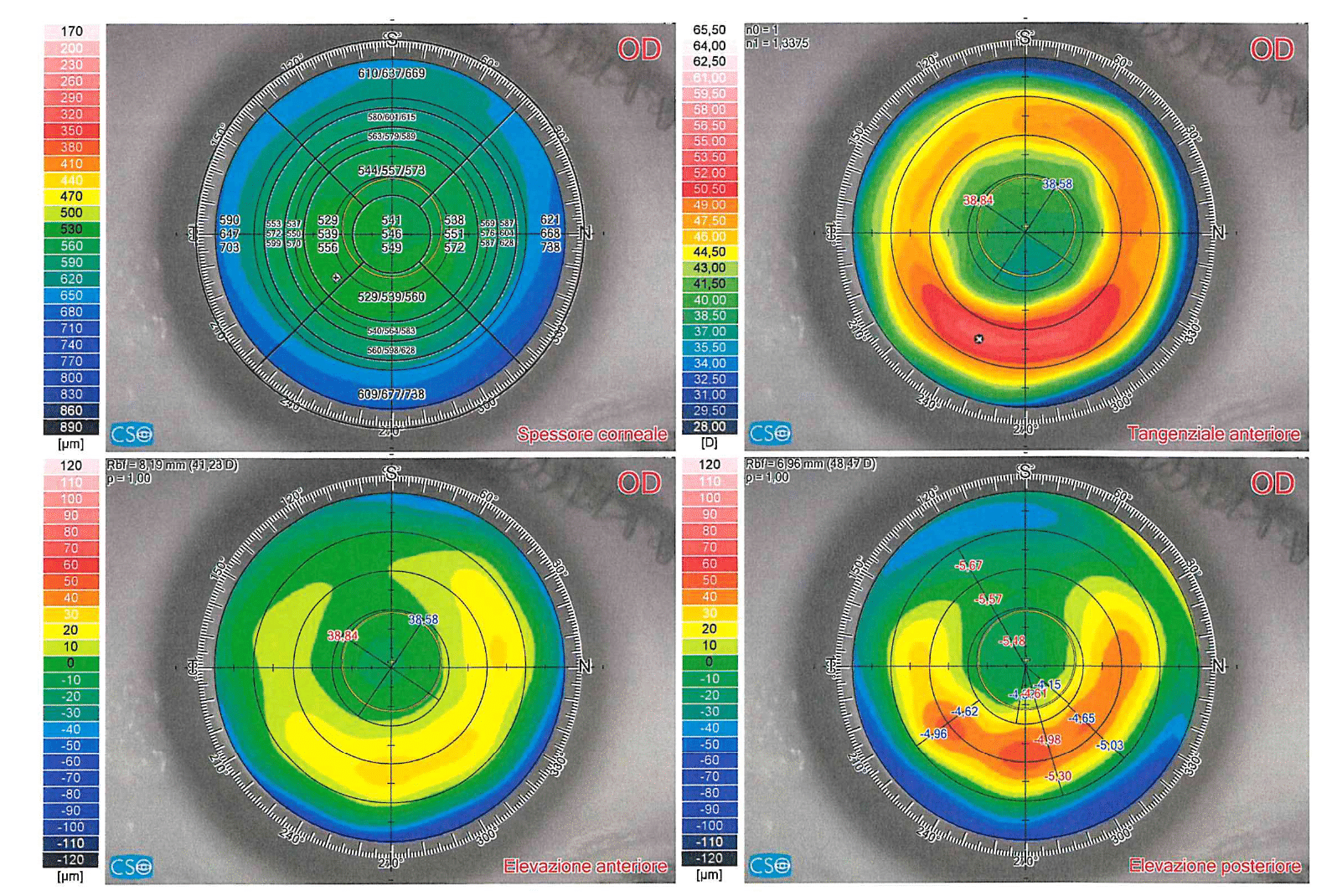
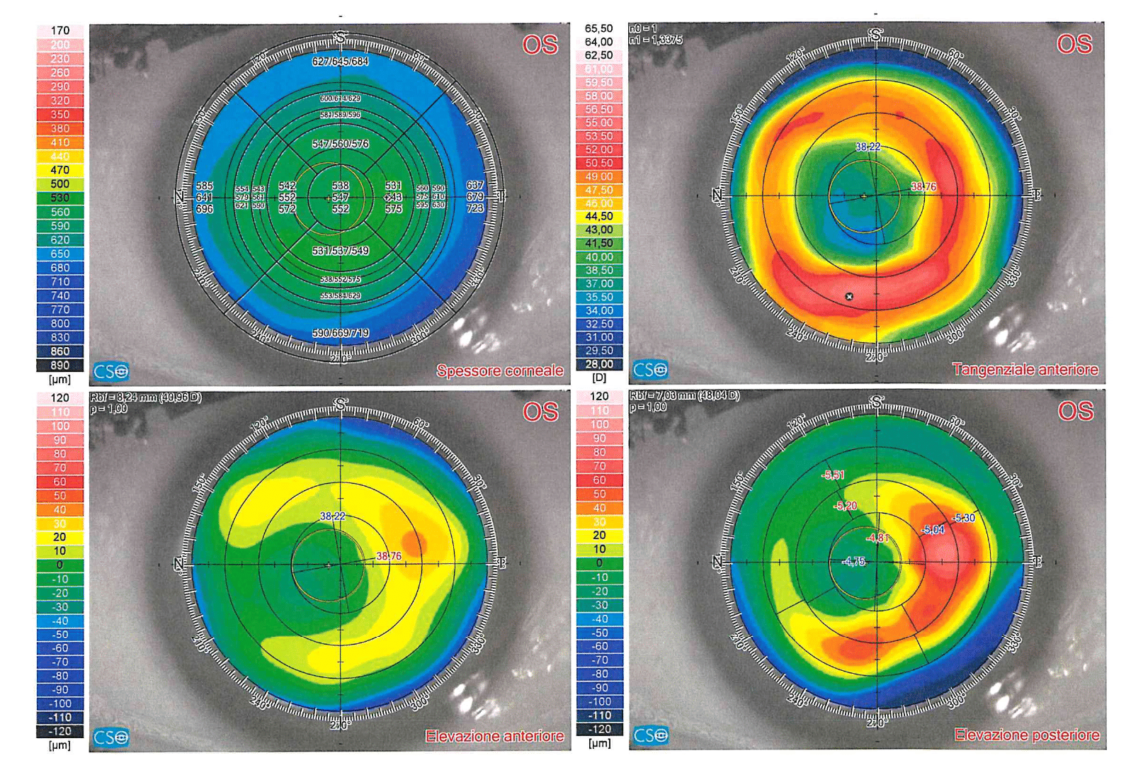
Radial keratotomy (RK) corrected myopia with deep radial incisions that weakened the corneal periphery. The periphery bulged, the centre flattened, myopia decreased. But a cornea cut to 90-100% of its thickness never recovers its rigidity. It goes on flattening for years, pushed by the intraocular pressure: in the PERK study, 43% of eyes shifted towards hyperopia by at least one dioptre over ten years, without ever reaching a plateau [1]. And the same cornea changes shape through the day: steeper in the morning, flatter in the evening, with a refractive fluctuation of about half a dioptre in symptomatic patients [2,3]. That a less rigid cornea is the cause has also been measured: in 64 eyes 24-32 years after RK, those with a hyperopic shift had lower corneal hysteresis and resistance factor, a central cornea more than 50 microns thinner, and a tendency to higher pressure than those that had stayed stable [11]. In these eyes the cornea, not the lamina cribrosa, has become the target of the pressure.
Cross-linking, developed for keratoconus, creates new bonds between the collagen fibres of the stroma and increases corneal rigidity. It is natural to think of it as a remedy for a cornea weakened by surgery. The problem is that in keratoconus cross-linking does something else as well: it flattens the cornea by about a dioptre. In keratoconus that is a benefit. After RK, where flattening is the very problem, it is the opposite.
What the literature says
Not much. No randomised trial, no peer-reviewed series of more than nine eyes, no follow-up beyond twelve months. Fewer than forty published eyes in total. I list them one by one, because it is the only honest way to present them.
- Mazzotta and colleagues, Siena, 2011 [4]. A single case: progressive ectasia with hyperopic shift ten years after RK, treated with an epithelium-off protocol. Uncorrected vision improved from 0.2 to 0.6 and corrected vision from 0.3 to 0.8 at twelve months. But this was a case of true ectasia, not of simple post-RK instability.
- Fuentes-Páez and colleagues, Barcelona, 2012 [5]. Seven eyes of four patients with refractive instability. At three months the mean sphere moved from −0.7 to +1.0 D, a statistically significant hyperopic shift. Visual acuity improved, but not significantly. Six months of follow-up.
- Elbaz and colleagues, Toronto, 2014 [6]. The most cited series: nine eyes of six patients who had RK 15 to 23 years earlier, treated for falling vision or fluctuation. In eight of nine eyes the diurnal fluctuation stopped between six and twelve months. But mean keratometry flattened by a dioptre, the spherical equivalent went from +1.4 to +2.5 D, and no change in acuity reached significance. The authors themselves note that part of the effect seen at six months had faded by twelve.
- Bafna, 2017 [7]. Fourteen eyes of seven patients treated with transepithelial cross-linking, published in a trade journal without peer review. Mean hyperopic shift of +0.72 D, mean flattening of 0.71 D, seven of fourteen eyes shifting by more than a dioptre. Patients who were already hyperopic reported seeing worse; the two still myopic, better. The author concludes by proposing cross-linking as a step before lens surgery, not as a treatment.
- Cubuk and Bilgihan, 2019 [8]. A negative case, and an atypical one: a 28-year-old woman with keratoconus treated by RK, a practice once used for keratoconus with frequently poor results. After cross-linking, corrected acuity fell from 0.9 to 0.6 and maximum curvature rose from 52 to 57 D in eight months. The most telling detail is where: the ectasia progressed exactly at two incisions with separated wound lips, visible at the slit lamp and on anterior segment OCT. The authors suggest that cross-linking may have stretched the incision margins, and state honestly that, having used different topographers before and after, they cannot separate the effect of the treatment from natural progression.
- Colombo-Barboza and colleagues, 2024 [9]. The most recent review on managing post-RK patients does not list cross-linking among the recommended options.
What emerges
Taken together, these papers say four things.
- On diurnal fluctuation, it works. This is the only consistent finding: in the Toronto series the fluctuation stopped in almost every eye. For the patient who sees well in the morning and poorly in the evening, or the reverse, that is a real benefit.
- On hyperopia, it makes it worse. In every series the cornea flattens by 0.7-1.1 dioptres after treatment. The typical post-RK patient is already hyperopic: cross-linking makes them more so. This is an expected consequence of the treatment’s biomechanics, not a surprise.
- On progression, we do not know. No study has followed patients long enough. The post-RK shift is measured in decades; the longest published follow-up is twelve months, and in that very series the effect was already regressing. To say that cross-linking “stops the shift” has, today, no basis.
- On safety, caution. No major complications in the small series, but one case of worsening and, in the transepithelial series, two eyes losing four lines to cataract. RK incisions, often gaping or plugged with epithelium, change the way riboflavin penetrates the stroma, and nobody has studied how.
Localised cross-linking: a different idea
There is a line of work that the Western literature never cites, and which I found only by searching in Russian. Anisimov’s group in Moscow turned the problem round: if standard cross-linking flattens because it stiffens the centre, then stiffen only the periphery. The technique removes the epithelium only over the incisions, applies riboflavin, and then irradiates through a mask cut to the patient’s own topography, exposing a ring over the incisions and shielding the central optical zone [13]. The aim is not to halt the shift but to reverse it, giving curvature back to the centre.
The published numbers are an order of magnitude larger than everything else: in a 2014 doctoral thesis, 105 eyes of 56 patients aged 48 to 53, with a mean hyperopia of +5.75 D, treated with this technique [12]. At one month, central keratometry is reported to have gone from 29 to 35 D, keratometric fluctuation to have halved, and corrected acuity to have risen from 0.08 to 0.90.
The reservations are just as large, and I set them all out. Follow-up is one month, against a shift measured in decades. The thesis was never published in an indexed journal, and the group’s peer-reviewed paper presents the method with one post-RK case, not the series [13]. A central steepening of six dioptres obtained by irradiation alone is an extraordinary claim, which no other group has replicated in twelve years. And yet the rationale fits what the Western literature suggests by another route: the weakness of a post-RK cornea lies in the incisions, and a treatment that acts on the incisions makes more sense than one that acts on the centre. It is an idea to follow, not yet a practice to adopt.
For completeness: two registered studies in the United States included or planned an arm for post-RK diurnal fluctuation, using transepithelial cross-linking [14,15]. Neither has published separate results for these eyes.
Why it may not be enough
There is also a structural reason why cross-linking struggles in this cornea, and it is worth explaining. Cross-linking stiffens the anterior stroma, the most superficial 250-300 microns, and acts on the zone that is irradiated. But the weakness of a post-RK cornea is not diffuse: it lies in the incisions, which cross almost the full thickness and run out to the limbus. Stiffening the surface between one incision and the next does not close the incisions. The cornea remains a structure of segments held together by scars, and the intraocular pressure keeps working on those scars. This is not only my reasoning: Mazzotta, in his case, already wrote that the effect of cross-linking does not reach the depth of the RK incisions [4], and in Cubuk’s case the ectasia progressed precisely where the incisions were open [8]. This explains, I believe, why the fluctuation settles while the slow shift does not stop.
When I offer it
In my practice, cross-linking after RK has a single, narrow indication: the patient with marked, disabling diurnal fluctuation, whose refraction changes too much through the day for an intraocular lens to be calculated reliably. In that case cross-linking can be a stabilising step. I wait at least six months for the refraction to settle, and then move on to lens surgery, with a lens calculated on a steadier cornea. The patient knows in advance that their hyperopia will have increased in the meantime.
I do not offer it as a treatment for hyperopia. Late post-RK hyperopia is corrected inside the eye, with a phakic ICL if the lens is clear and the anterior chamber allows it, or with lens replacement and a small-aperture lens if it does not. I discuss this in a separate article. And I do not offer it to the patient whose fluctuation is modest: there the benefit is uncertain and the price, an extra dioptre of hyperopia, is certain.
For completeness: concentric corneal sutures also exist, which steepen the centre and reduce hyperopia, with results documented in small series [10]. They work, but they regress and need managing over time. Those too I reserve for selected cases.
What this changes in practice
Cross-linking after radial keratotomy is not a solution for late hyperopia. It is a tool for stabilising a fluctuating cornea before the refraction is corrected by other means. Whoever offers it as a cure should show data that, in the verifiable literature, do not yet exist. Whoever dismisses it altogether gives up the one effect the literature supports, and ignores an idea, localised treatment over the incisions, that deserves proper study. The right path lies between: use it for what it does, and tell the patient beforehand. And, for these patients, measure the pressure with non-applanation methods and keep them under glaucoma surveillance: the same weak cornea that flattens is also a cornea that makes tonometry unreliable.
The information in this article is for general guidance and does not replace an eye examination. Every treatment decision should follow a complete assessment of the eye.
References
- Waring GO 3rd, Lynn MJ, McDonnell PJ; PERK Study Group. Results of the Prospective Evaluation of Radial Keratotomy (PERK) Study 10 years after surgery. Arch Ophthalmol. 1994;112(10):1298-1308. doi:10.1001/archopht.1994.01090220048022
- McDonnell PJ, Nizam A, Lynn MJ, Waring GO 3rd; PERK Study Group. Morning-to-evening change in refraction, corneal curvature, and visual acuity 11 years after radial keratotomy in the Prospective Evaluation of Radial Keratotomy Study. Ophthalmology. 1996;103(2):233-239. doi:10.1016/s0161-6420(96)30711-2
- Kemp JR, Martinez CE, Klyce SD, et al. Diurnal fluctuations in corneal topography 10 years after radial keratotomy in the Prospective Evaluation of Radial Keratotomy Study. J Cataract Refract Surg. 1999;25(7):904-910. doi:10.1016/s0886-3350(99)00090-5
- Mazzotta C, Baiocchi S, Denaro R, Tosi GM, Caporossi T. Corneal collagen cross-linking to stop corneal ectasia exacerbated by radial keratotomy. Cornea. 2011;30(2):225-228. doi:10.1097/ICO.0b013e3181e16de5
- Fuentes-Páez G, Castanera F, Gómez de Salazar-Martinez R, et al. Corneal cross-linking in patients with radial keratotomy: short-term follow-up. Cornea. 2012;31(3):232-235. doi:10.1097/ICO.0b013e31821f28bb
- Elbaz U, Yeung SN, Ziai S, et al. Collagen crosslinking after radial keratotomy. Cornea. 2014;33(2):131-136. doi:10.1097/ICO.0000000000000044
- Bafna S. Can cross-linking help after radial keratotomy? Cataract & Refractive Surgery Today. July 2017. crstoday.com
- Cubuk MO, Bilgihan K. Performing corneal collagen cross-linking after radial keratotomy in a patient with keratoconus: case report. Beyoglu Eye J. 2019;4(3):206-209. doi:10.14744/bej.2019.79653
- Colombo-Barboza GN, Rodrigues PF, Colombo-Barboza FDP, et al. Radial keratotomy: background and how to manage these patients nowadays. BMC Ophthalmol. 2024;24(1):9. doi:10.1186/s12886-023-03261-0
- Nosé W, Endriss D, Forseto AS. Corneal suture for the correction of hyperopia following radial keratotomy. J Refract Surg. 2007;23(5):523-527. doi:10.3928/1081-597X-20070501-19
- Avetisov SE, Antonov AA, Vostrukhin SV. Progressive hyperopic shift after radial keratotomy: possible causes. Vestn Oftalmol. 2015;131(2):13-18 (in Russian). doi:10.17116/oftalma2015131213-18
- Smotrich EA. Corneal topography and distribution of mechanical stress in different types of corneal surgery. PhD thesis (supervisor SI Anisimov), Institute for Advanced Medical Training, Federal Medical-Biological Agency, Moscow, 2014 (in Russian). www.mntk.ru
- Anisimov SI, Anisimova SY, Mistryukov AS. Personalized (local) UV-crosslinking as a treatment of keratoconus and corneal ectasia. Ophthalmology in Russia. 2017;14(3):195-199 (in Russian). doi:10.18008/1816-5095-2017-3-195-199
- ClinicalTrials.gov NCT02095730. Corneal cross-linking comparing variables (arm: RK/AK diurnal fluctuation). clinicaltrials.gov
- ClinicalTrials.gov NCT03029104. Collagen cross-linking with ultraviolet-A in asymmetric corneas (CXLUSA; includes diurnal fluctuation after radial keratotomy). clinicaltrials.gov

