



Abstract
Purpose: To investigate the dimensional changes that occur in the polymethylmethacrylate (PMMA) haptics of three-piece intraocular lenses (IOLs) after prolonged implantation and their implications for the Yamane technique of intrascleral fixation.
Methods: Review of the scientific literature on the biochemical and structural changes of PMMA haptics, comparative analysis of the biomechanical properties of different three-piece IOLs, and evaluation of the clinical impact on the Yamane technique.
Results: PMMA haptics undergo multifactorial dimensional changes over time, including protein deposition (5-15 µm), capsular fibrosis with mechanical compression (5-10 µm), water absorption (5-10 µm) and plastic deformation (5-10 µm). The total dimensional increase of 20-50 µm can compromise the passage through 30G thin-wall needles (internal diameter 0.159-0.17 mm) used in the Yamane technique. IOLs with PVDF haptics demonstrate superior biomechanical stability compared to PMMA.
Conclusions: Knowledge of the dimensional changes of the haptics is essential for surgical planning in the Yamane technique, especially when using explanted IOLs. The appropriate selection of the IOL and the needle gauge is fundamental to guarantee long-term stability.
Keywords: Yamane technique, PMMA, three-piece IOL, intrascleral fixation, haptics, dimensional changes
1. Introduction
Intrascleral fixation of intraocular lenses (IOLs) represents a fundamental solution in the absence of capsular support, a condition that occurs in approximately 5-10% of cataract surgeries due to intraoperative complications, trauma, or late dislocation of the IOL. [1,2]
The intrascleral fixation technique with haptic flanges, originally described by Shin-ichi Yamane and colleagues in 2017, has revolutionized the surgical approach by eliminating the need for sutures, reducing operative time, and minimizing long-term complications associated with suture degradation. [3]
The original Yamane technique uses 30-gauge (30G) thin-wall needles to create transconjunctival scleral tunnels through which the haptics of three-piece IOLs are externalized. The end of the haptics is then cauterized to create a “flange” which, once repositioned in the scleral tunnel, prevents the dislocation of the IOL. [3,4]
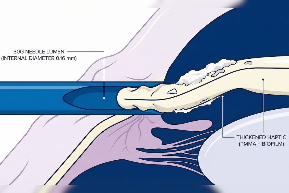
However, clinical experience has highlighted that the haptics of explanted IOLs, having remained in the eye for several years, show a significantly greater resistance to passage through 30G needles compared to the haptics of new IOLs. This phenomenon raises questions about the structural and dimensional changes that occur in PMMA haptics over time.
1.1 Purpose of the Study
- Analyze the biochemical and biomechanical mechanisms responsible for the dimensional changes of PMMA haptics
- Quantify the extent of these changes based on the available literature
- Evaluate the clinical implications for the Yamane technique
- Provide evidence-based recommendations for surgical management
2. Materials and Review Methodology
A systematic review of the literature was conducted using the PubMed, Scopus, Web of Science, and Cochrane Library databases. Studies published between 2000 and 2025 were included.
3. Anatomy and Characteristics of Three-Piece IOLs
3.1 Haptic Materials
3.1.1 PMMA (Polymethylmethacrylate)
- AcrySof MA60AC (Alcon): haptic diameter 0.150 ± 0.005 mm
- Sensar AR40E (J&J): haptic diameter 0.146 mm
- Tecnis ZA9003 (J&J): haptic diameter 0.146 ± 0.004 mm
3.1.2 PVDF (Polyvinylidene Fluoride)
- CT Lucia 602 (Zeiss): haptic diameter 0.147 ± 0.005 mm. Offers greater tensile strength and formation of more stable flanges.
3.2 Critical Dimensional Parameters
| Parameter | Dimension (mm) |
|---|---|
| 30G thin-wall needle – Inner diameter | 0.159 – 0.192 |
| PMMA haptic (MA60AC) – unmodified | 0.150 ± 0.005 |
| PVDF haptic (CT Lucia) – unmodified | 0.147 ± 0.005 |
| Tolerance margin | 0.009 – 0.042 |
4. Mechanisms of Dimensional Modification of the Haptics
4.1 Protein Deposition and Biofilm
Microscopic studies on explanted IOLs have detected a protein layer (fibronectin, albumin) of variable thickness between 5-15 µm on the haptics.
4.2 Capsular Fibrosis
Capsular contraction exerts mechanical forces that can cause a plastic deformation of the haptic section, with an increase in the maximum diameter of 5-10 µm.
4.3 Water Absorption
Despite its hydrophobicity, PMMA can absorb up to 2-3% of water in saturation conditions after years, leading to an estimated expansion of 5-10 µm.
4.5 Plastic Deformation from Chronic Stress
Under the constant load of the contracted capsule, PMMA (viscoelastic material) undergoes a “creep” process. The formula that describes such deformation over time is:
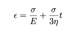
This involves a radial dimensional increase of approximately 5-10 µm after 5 years.
5. Quantitative Synthesis: Total Dimensional Increase
| Mechanism | Increment (µm) |
|---|---|
| Protein deposition and biofilm | 5 – 15 |
| Peri-haptic capsular fibrosis | 5 – 10 |
| Water absorption/swelling | 5 – 10 |
| Superficial calcification | 2 – 10 |
| Plastic deformation | 5 – 10 |
| TOTAL | 22 – 55 |
Result: The total dimensional increase (22-55 µm) often exceeds the tolerance margin of the 30G needle (9-42 µm), making the passage of the explanted haptic extremely difficult.
6. The Yamane Technique: Biomechanical Aspects
Studies demonstrate that the dislocation force with a 30G needle is decidedly superior for PVDF (2.04 N) compared to PMMA (0.27 – 0.93 N). A correlation between the flange/needle ratio and dislocation force has been identified (r=0.975, p < 0.001).
9. Conclusions
The present study confirms that PMMA haptics undergo a volumetric increase over time. For primary interventions, it is preferable to use IOLs with PVDF haptics. For revisions of explanted IOLs, the surgeon must consider the use of 27G needles or lens replacement if dimensional modifications prevent safe externalization.

